Digipath AI Test

PRECISION SURGERY WITH VELSCOPE Vx: ​

Revolutionary Fluorescence-Guided Surgery for Oral Cancer VELscope Vx represents a paradigm shift in oral cancer surgery, utilizing tissue fluorescence visualization (TFV) to identify malignant tissue invisible to the naked eye. This breakthrough technology enables surgeons to achieve unprecedented precision in tumor removal, dramatically reducing recurrence rates and improving patient outcomes.

The Challenge: Limitations of Conventional Surgery

Traditional White Light Surgery Falls Short

Conventional oral cancer surgery relies on visual assessment under white light, which presents significant limitations:

Inadequate Visualization: Tumor boundaries are often unclear under standard lighting

Incomplete removal: Microscopic cancer cells may remain at surgical sites

Guesswork margins: Surgeons must estimate safe surgical margins without precise guidance

High recurrence rates: Up to 40% of patients experience cancer recurrence after conventional surgery

The Cost of Imprecision

When surgical margins are inadequate, the consequences are severe:

Cancer recurrence: Remaining malignant cells lead to tumor regrowth

Additional surgeries: Patients require repeat procedures with increased complexity

Reduced survival rates: Recurrent cancer is typically more aggressive and harder to treat

Escalated costs: Second surgeries, chemotherapy, and radiation multiply treatment expenses

VELscope Vx: The Solution for Precision Surgery

VELscope Vx utilizes a specific wavelength of blue light to reveal tissue abnormalities:

Blue light excitation: The device emits 400 – 460nm wavelength light

Cancer detection: Abnormal tissue shows loss of fluorescence and appears dark

Normal tissue response: Healthy tissue fluoresces and appears as an apple-green glow

Real-time guidance: Surgeons can visualize the exact extent of cancerous tissue during surgery

The Science Behind Fluorescence Loss

Normal Tissue Fluorescence:

Healthy epithelium contains fluorophores that emit green light

Intact collagen matrix in the stroma maintains fluorescence

Blood vessels and normal architecture support fluorescence

Cancer-Associated Changes:

Epithelial dysplasia disrupts normal fluorophore distribution

Collagen matrix breakdown reduces fluorescence intensity

Increased blood vessel density and altered tissue architecture cause fluorescence loss

Clinical Evidence: Dramatic Improvement in Outcomes Proven Recurrence Reduction

Clinical studies demonstrate the remarkable effectiveness of VELscope-guided surgery:

Squamous Cell Carcinoma Study:

92 patients with SCC: 3-year local recurrence rate reduced from 40.6% to 6.5%

Over 80% reduction: Fluorescence guidance dramatically improved outcomes

High-Grade Lesions Study:

62 patients with severe dysplasia: Recurrence rate decreased from 39.3% to 8.1%

Consistent results: Benefits observed across different lesion types

Long-Term Follow-Up Results

Non-Fluorescence Guided Surgery:

25% of patients experienced severe dysplasia or worse at
treated sites after 12 months

Higher rates of positive margins requiring additional treatment

VELscope-Guided Surgery:

0% of patients had severe dysplasia at treated sites

Complete removal achieved in significantly more cases

Benefits of VELscope-Guided Surgery

Immediate Surgical Benefits

Precise tumor delineation: Clear visualization of cancer boundaries

Complete removal: Ensures no malignant tissue remains

Reduced operative time: Real-time guidance streamlines procedures

Improved margins: Higher rates of clear surgical margins

Long-Term Patient Benefits

Dramatically reduced recurrence: Over 90% reduction in local recurrence rates

Enhanced survival: Complete tumor removal improves long-term prognosis

Fewer repeat surgeries: Reduced need for additional procedures

Better quality of life: Successful single surgery minimizes patient trauma

Healthcare System Benefits

Cost reduction: Preventing recurrence saves substantial healthcare resources

Improved efficiency: Precise surgery reduces complications and readmissions

Enhanced outcomes: Superior results improve institutional reputation

Resource optimization: Better allocation of surgical and treatment resources

​PRECISION SURGERY WITH VELSCOPE Vx: ​ REDUCING ORAL CANCER RECURRENCE BY UP TO 90%​

Initial assessment of an ill-defined SCC on the right ventrolateral tongue under white light​.

Clinically apparent tumor outlined in blue​.

Assessment of field using FV in the dark​.

FVL area outlined in green, with the boundary of the surgical specimen marked in red.​

What experts have to say:

“We see recurrence rate decrease significantly in both, invasive cancers and cases of severe dysplasia, when surgical margins are determined using Tissue Fluores-cence Visualization.”

Dr. Calum MacAulay PhD Clinical Associate Professor, UBC
Head of Integrative Oncology Distinguished Scientist Serves as Head, Cancer Imaging Department, B.C. Cancer Research Centre, BCAA

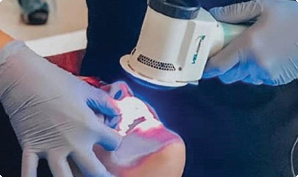
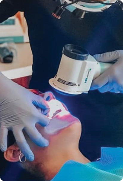
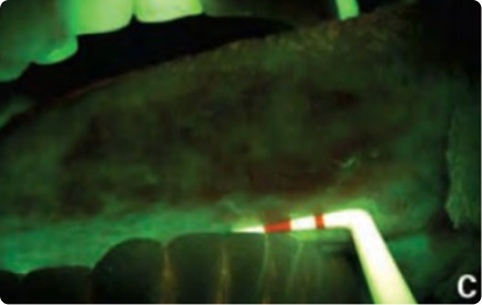
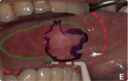
“With FV technology we found that the extension of this disease is not uniform around the cancerous area, so a 10-mm margin around the tumor site may not always work. With the use of FV technology, we hope to accurately remove all cancerous tissue.”

Prof. Dr. Catherine F. Poh, DDS, PhD, FRCD (C), Clinician Scientist, Oral Pathologist and Consulting Dentist, BC, Canada.

“VELscope allows us to add new dimension to detect abnormalities which may be histologic or perhaps only genetic and use those in real time way to evaluate surgical margins”

Prof. J. Scott Durham, BSc MD FRCSC Head, VGH Otolaryngology – Head and Neck Surgery