Digipath AI Test

Eliminate Biopsy Errors

Biopsy sampling errors are a critical challenge in oral cancer diagnosis, leading to delayed treatment, missed diagnoses, and poor patient outcomes. Traditional white light examination makes it difficult to pinpoint the exact location of malignant tissue, resulting in inaccurate sampling and compromised diagnostic accuracy. However, advanced fluorescence visualization technology now offers a revolutionary solution to eliminate these errors entirely.

The Critical Problem: Biopsy Sampling Errors

High Error Rates Under White Light

Traditional biopsy procedures face significant limitations that compromise diagnostic accuracy

60% discordance rate: Sampling errors occur in 60% of cases due to inadequate site identification

Invisible lesions: 11.1% of oral lesions are missed under white light examination

Guesswork-based sampling: Clinicians must rely on visual estimation to determine biopsy sites

Extensive lesion areas: Large leukoplakic patches make identifying precise biopsy locations extremely challenging

Consequences of Sampling Errors

When biopsy sites are incorrectly identified, the consequences can be devastating:

Delayed diagnosis: Sampling from non-malignant areas leads to false-negative results

Repeated procedures: Patients require multiple biopsies, increasing discomfort and costs

Disease progression: Missed diagnoses allow cancer to advance to later stages

Reduced survival rates: Late-stage detection dramatically decreases treatment success

Revolutionary Solution: VELscope Vx Fluorescence Guidance Precise Lesion Identification

VELscope Vx utilizes tissue fluorescence visualization to reveal abnormalities invisible to the naked eye:

How it works:

Blue light excitation: The device emits 400 – 460nm wavelength light

Abnormal tissue detection: Malignant areas show loss of fluorescence (LOF) and appear dark

Normal tissue response: Healthy tissue fluoresces and appears as an apple-green glow

Real-time visualization: Clinicians can immediately identify exact biopsy sites

Eliminating Sampling Errors

Before Velscope Guidance:

With VELscope Guidance:

High risk of sampling from non-malignant tissue

Loss of fluorescence highlights areas of concern: Two distinct dark regions clearly visible under fluorescence

Clinicians must estimate the most suspicious locations

Precise targeting: Biopsy sites are definitively identified based on fluorescence patterns

Extensive leukoplakic areas under white light make precise site identification impossible

Zero discordance: Sampling errors are eliminated through accurate visualization

Clinical Evidence: Proven Error Elimination Dramatic Improvement in Accuracy

Clinical studies demonstrate the remarkable effectiveness of fluorescence-guided biopsy:

Traditional White Light Biopsy:

  • 60% discordance rate: High rate of sampling errors due to inadequate visualization
  • Missed diagnoses: Significant number of malignant lesions go undetected
  • Repeated procedures: Patients require multiple biopsies to achieve accurate diagnosis

VELscope-Guided Biopsy:

  • Zero discordance rate: Fluorescence guidance eliminates sampling errors
  • Accurate targeting: Malignant tissue is precisely identified and sampled
  • Single procedure success: Correct diagnosis achieved in first biopsy attempt

Enhanced Diagnostic Confidence

The integration of fluorescence visualization with traditional examination provides:

Objective guidance: Removes subjective guesswork from site selection

Visual confirmation: Clinicians can see exactly where abnormal tissue is located

Reproducible results: Consistent accuracy across different practitioners

Immediate feedback: Real-time visualization during procedure

ELIMINATE ORAL BIOPSY SAMPLING ERRORS: ESSENTIAL FOR ACCURATE DIAGNOSIS AND EFFECTIVE TREATMENT PLANNING​

PRECISE BIOPSY SITES ARE CHALLENGING TO IDENTIFY UNDER WHITE LIGHT​

60% Discordance due to sampling errors

The extensive area of leukoplakia under white light makes identifying the exact biopsy site challenging.​

Fluorescence visualization helps in identifying appropriate sites for biopsy

Biopsy Discordance rate goes to zero

Loss of fluorescence highlights areas of concern (two distinct dark regions as observed under VELscope). ​
Biopsy of these areas revealed moderate dysplasia.​

Benefits of Eliminating Biopsy Errors

For Patients

Reduced trauma: Single accurate biopsy eliminates need for repeat procedures

Faster diagnosis: Correct results obtained immediately

Lower costs: Eliminates expenses associated with multiple biopsies

Earlier treatment: Accurate diagnosis enables timely intervention

For Healthcare Providers

Improved accuracy: Confidence in diagnostic results

Enhanced reputation: Superior diagnostic capabilities

Reduced liability: Minimized risk of missed diagnoses

Streamlined workflow: Efficient single-procedure approach

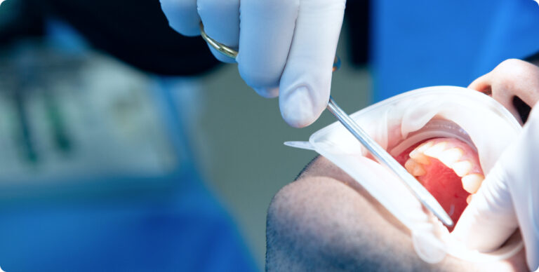
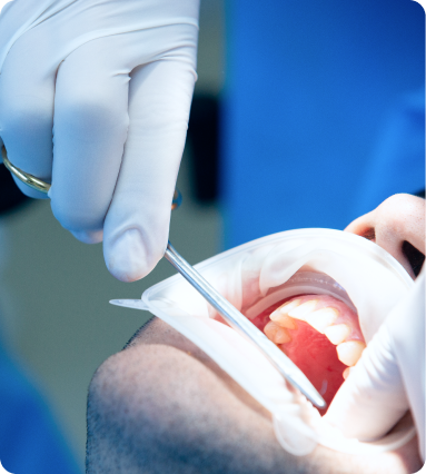
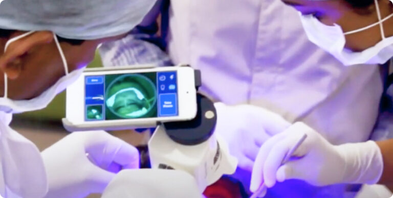
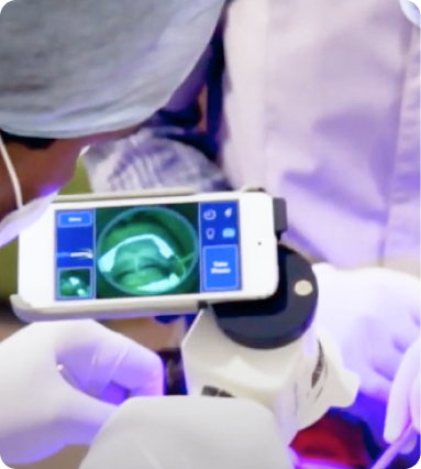
Biopsy sampling errors are a preventable cause of diagnostic delays and missed cancers. With VELscope Vx fluorescence visualization, healthcare providers can eliminate these errors entirely, ensuring accurate diagnosis on the first attempt. This technology represents a fundamental shift from uncertainty to precision, from
repeated procedures to single-attempt success.

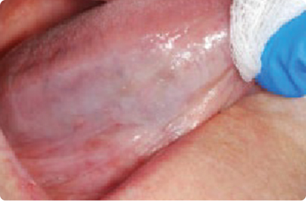
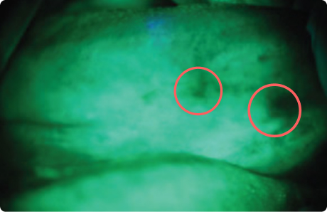
The evidence is clear: Fluorescence-guided biopsy eliminates sampling errors and dramatically improves diagnostic accuracy. Every patient deserves the precision and confidence that comes with advanced visualization technology.

The power to eliminate biopsy errors is available today. The question is not whether this technology works—clinical evidence provides definitive proof. The question is whether healthcare providers will embrace this advancement to improve patient outcomes.